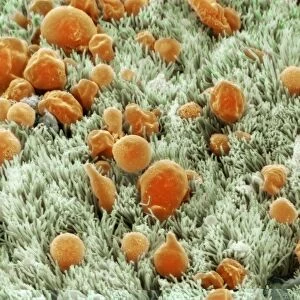
Trachea lining, SEM C015 / 9939

![]()
Wall Art and Photo Gifts from Science Photo Library
Trachea lining, SEM C015 / 9939
Trachea lining. Coloured scanning electron micrograph (SEM) of a section through the lining of the trachea (wind pipe). The trachea links the larynx (voice box) to the lungs. Pseudostratified ciliated columnar epithelial cells, which line the trachea, are bright orange. The hair-like cilia (yellow) on their surface beat to move mucous, and particles trapped in it, upwards out of the respiratory tract. This helps to keep the lungs and airways clear and prevent infection. At bottom is the basement membrane, a layer of connective tissue that supports the epithelial cells
Science Photo Library features Science and Medical images including photos and illustrations
Media ID 9239465
© SUSUMU NISHINAGA/SCIENCE PHOTO LIBRARY
Basement Membrane Cilia Columnar Epithelium Connective Tissue Epithelial Epithelium Lining Respiratory Scanning Microscopy Trachea Tract Wind Pipe Cells Section Sectioned
EDITORS COMMENTS
This print showcases the intricate beauty of the trachea lining, as captured through a scanning electron microscope (SEM). The vibrant colors bring to life the complex structure and function of this vital respiratory organ. The trachea, also known as the windpipe, acts as a crucial conduit between the larynx and lungs. In this image, we can observe pseudostratified ciliated columnar epithelial cells that line the trachea. These specialized cells are depicted in bright orange hues, creating a striking contrast against their surroundings. What truly stands out in this image are the hair-like structures called cilia that adorn the surface of these epithelial cells. Colored in yellow, these tiny cilia play an essential role in maintaining respiratory health. They beat rhythmically to propel mucus and any trapped particles upwards and out of our airways. This process helps keep our lungs clear from potential infections. At the bottom of this microscopic view lies another important component - the basement membrane. Composed of connective tissue, it provides support for these delicate epithelial cells. Through SUSUMU NISHINAGA's expert lens and advanced microscopy techniques, we gain a deeper appreciation for both the complexity and elegance found within our own bodies. This mesmerizing photograph serves as a testament to science's ability to unveil hidden wonders at even microscopic scales.
MADE IN THE USA
Safe Shipping with 30 Day Money Back Guarantee
FREE PERSONALISATION*
We are proud to offer a range of customisation features including Personalised Captions, Color Filters and Picture Zoom Tools
SECURE PAYMENTS
We happily accept a wide range of payment options so you can pay for the things you need in the way that is most convenient for you
* Options may vary by product and licensing agreement. Zoomed Pictures can be adjusted in the Cart.